


Osteocondrosis of the lumbar region is a chronic degenerative disease of the lumbar column that affects the structure of the intervertebral discs and a series of lumbar vertebrae positioned.It affects people of the mainly working age.It manifests itself in various symptoms, whose main is pain in the lower back and legs, limiting the movements in the lower back.For diagnostics, research methods such as radiography, computerized tomography or magnetic resonance imaging of the lumbar column are used.In this article, you can more detail with the causes, symptoms and methods to diagnose the osteochondosis of the lumbar column.
Osteocondrosis is the result of body aging.These or other signs of this disease can be found in almost all people (!), Starting from 25 years.But here is the seriousness of these changes, the rate of their progression, the degree of clinical manifestations depends on many causes, mainly on how a healthy lifestyle leads a specific person.Moderate physical activity, compulsory morning gymnastics, the right installation of the body during the execution of a series of works (garden, construction, trivial cleaning of the house and so on), the orthopedic mattress is those moments that prevent the development of the osteochondosis of the lumbar column.
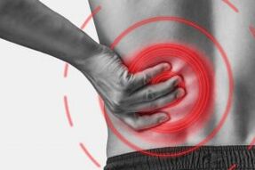
According to statistics, the osteochondosis of the spine in 80% of cases is the cause of back pain.
How does osteochondrosis develop?
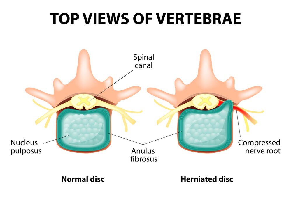
The entire spine consists of separate vertebrae, among the bodies of which there are intervertebral discs.That is, between the two vertebrae there is a disc.The disc consists of a gelatinous nucleus (pulpic) and a fibrous ring.The nucleus contains a lot of water and provides amortization and flexibility of the spine.The fibrous ring is located along the outskirts of the nucleus of the jacket, as if he kept it inside himself.
With a prolonged increased load on the core of the pier, it changes its physiological properties, loses water and drying and finally sequences: the disc is flattened and the vertebral bodies approach each other.Together with these processes, in the nucleus jacket, the fibrous ring loses its elasticity and, under the influence of mechanical loads, begins to protrude.This is called protrusion.Then the fibrous ring breaks and a gelatinous nucleus falls through the resulting spaces: a disc of the disc occurs.A diagram of two adjacent vertebrae and a disc located with each other, called vertebral segment, acquires excess mobility, thus increasing the load on the neighboring segments.The overload of nearby segments triggers a similar pathological process in them.These changes are called osteochondrosis.
In order to somehow guarantee the stability of the spine, the bone growths are formed along the edges of the vertebral bodies, increasing the support area.This phenomenon is called spondylosis.The changes in the joints between the vertebrae are called arthrosis sponder.Usually all three pathologies - osteochondrosis, spondylosis, spondic arthrosis - walk nearby.
Reasons
Why does osteochondrosis occur?To date, there are several theories of occurrence:
- Mechanical theory: perhaps the main reason should be considered a regular load on the spine.That's why osteochondosis is an almost mandatory destiny of removals, miners, manufacturers and people of these professions.The presence of osteochondosis of the lumbar region is mainly associated with the slopes and the lifting of gravity, forced an uncomfortable laying of work;
- Another factor in development is the incorrect posture, sitting in the wrong laying, which is particularly relevant for mental workers;
- Sometimes the role is played by the hereditary characteristics of the structure of the spine and the nutrition of its individual structures;
- Traumatic theory: any trauma to the spine (even the most insignificant) is able to launch a degenerative process;
- Hormonal metabolic disorders and endocrine diseases can negatively influence the metabolism in the tissues of the spinal column and contribute to the development of osteocondrosis;
- Age theory implies the natural wear of records in the life process.
Rarely, only one of these theories can explain the occurrence of osteochondrosis in each case.More often at the same time, several factors are "to blame".
In the occurrence of the osteochondosis of the lumbar column, overweight plays an important role, since itself is an overload for the spinal column.The greater the body mass index (degree of obesity), the more changes in the spine are usually pronounced.Among the other reasons that cause the appearance of osteochondrosis, it can be seen:
- sedentary lifestyle;
- Imorly nutrition (fast food, sweet excess products, semifinites: all this leads to an imbalance of trace elements) and a lack of liquid;
- anomalies of the structure of the spine (for example, the presence of a further lumbar vertebra);
- Wearing constant of high wheeled shoes;
- pregnancy (due to the excess load on the lumbar column);
- sudden interruption of training in people involved professionally in sport;
- Smoking and alcohol abuse: as factors that accelerate the aging process in the body.
Symptoms
The main event of the osteochondosis of the lumbar column is pain.The nature of the pain, the place of the occurrence and the direction of the distribution depend on which receptors are irritated, that is, how rough changes in the disc and in the surrounding tissues there are protrusion or hernia, in what direction the protrusion has been formed and so on.
Reflex and compression syndromes are distinguished with osteochondrosis of the lumbar column.
The syndromes reflects are developed in cases where the fibrous receptors of the disc, the ligaments and the joint capsules located nearby are irritated.They are reflective because in addition to the pains they are accompanied by changes of muscle-syical, vegetative-vascular or neurodist reflection, that is, the irritation with the reflections is transmitted to other structures, causing symptoms mainly from the side of the soft tissues.
The compression syndromes occur following the compression (compression) of the nerve roots, of the blood vessels or the spinal cord formed by the osteocondrosis by changes.

Reflex syndromes of the lumbar column
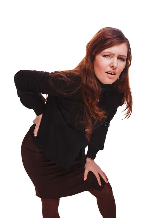
Lumbago(Feeling): Sudden acute pain in the lower back, which occurs with an embarrassing movement or at the time of physical tension (much less often - without an apparent reason).It is believed that the occurrence of Lumbago is associated with the movement of a nucleus of the jacket inside the fibrous ring, that is, it develops in the initial stages of the osteocondrosis.Often the pain is described as "feeling", "the pole was blocked in the lower back."Patients freeze in the installation in which the pain has captured them.The slightest move causes an increase in pain (sneezing, cough, an attempt to turn to bed, to move the foot).If a person was in an inclined position at the time of the development of Lumbago (which happens most often), then it cannot be straightened.A pronounced muscle tension in the lumbar column occurs reflectively.Along the vertebrae in this area, a muscle roller is warned, which is sometimes visible to the naked eye without touch, and muscle tension is thus pronounced.Feel painful for the patient.Such an increase in muscle tone plays a immobilizing role, protecting the lumbar segment affected by pathological mobility, which can cause a deterioration of the state.The natural curve of the spine in the lower back (lordosis) is flattened, perhaps the curvature (scoliosis) is possible due to muscle tension.
Lumagly- Another reflected syndrome of the lumbar level.This term also means the presence of pain in the lumbar region.But, unlike Lumbago, the pain does not show up, but gradually, in a few hours or even days.The pain is stupid, a moderate intensity, intensifies during the movements, in a sitting or standing position, when it moves from one position to another.A little relief brings the deck position or the back with a roller in the lower back of the back, but the passive rise of the leg straightened in this position causes an increase in pain in the lower back (symptom of lassa).The palpation of the lumbar column is painful, but the reflected tension of the muscles is less pronounced than lumbago, and sometimes absent.The movements in the lumbar column are limited, but possible.This means that the patient can bend and on the sides at a certain level (and therefore the pain intensifies).
Sciatica- Another variety of reflex syndrome of the lumbar level.This term means pain in the lower back, which gives the buttock and in the leg (on the rear surface).The pain is different, mostly painful, but it can periodically intensify from the type of "fireplace" in the leg.Just like with low back pain, it intensifies with any movements, walking, striving, decreasing in the back on the back.The symptom of lassa is generally positive.The palpation of the lumbar column is painful, in addition to pressing on some points (for example, in the middle of the line that separates the buttock from the thigh, in the middle of the back of the thigh, in the middle of the popliteal pit).There is tension of the muscles of the lower back.The inclinations forward and on the sides are limited.
Compression syndromes of the lumbar column
The clinical feature depends on which structure is subject to compression.
Among the vertebrae in each intervertebral hole there are nerve roots (spinal nerves): left and right.If the pathological formations for the osteochondosis of the lumbar column (mainly discs of the discs) squeeze the roots, the radicolopathy develops, whose symptoms differ for each root.Common to all the radicolopathies of the lumbar region is the increase in pain during sneezing, coughing, movement in the lower back (in particular inclining forward), the presence of muscle tension in the lower back of the back, the limitation of movements in the lumbar column.The following types of lumbar column radicolopathies are more common:
- Radicolopathy L1, L2, L3: the pain occurs in the lower part of the back, gives the expected thigh.In the same area, it is possible to occur from the paresthesia (a sense of gemito uvallo, numbness), the surface sensitivity is disturbed (a sharp touch from the usual does not stand out, the feeling of cold and hot) is lost.The reflection of the knee is revealed, the weakness of the quadriceps of the thigh;
- Radicolopathy L4: the pain from the lower back gives the front of the thigh, the internal surface of the knee joint and slightly lower along the internal surface of the lower part of the leg.In the same areas we feel the paresthesia and the surface sensitivity is lost (reduced).Weakness also develops in the muscle of the thigh quadriceps, the reflection of the knee decreases;
- Radicolopathy L5: one of the frequent locations.The pain gives the buttock, along the outer edge of the thigh, along the front surface of the lower leg to the internal edge of the foot and thumb.The paresthesia is felt here, the surface sensitivity is disturbed and pain is given here when it sneezes and coughs.In addition, there is a difficulty in extension of the thumb of the foot, since the muscle that performs this action is innervated by the Kine L5.Sometimes it is difficult to stay on a heel with a footed foot;
- Radicolopathy S1 is often also found with osteochondrosis of the lumbar column.The pain gives the buttock, along the outer edge of the thigh, along the outer edge of the lower leg to the outer edge of the foot and the 5th finger, the heels.These areas are characterized by a feeling of walls, a decrease in surface sensitivity.Achille's reflection is reduced.With the damage to this vertebral column, the weakness of the muscles of the lower part of the leg and develops the flexors of the foot, so standing and walking on the socks are difficult.
The simultaneous development of radicolopathies of different roots is possible, this is particularly characteristic of L5, S1.It happens that a hernia holds several roots.
If the herd of the disc attacks, then it can squeeze the spinal cord.This is possible only when the hernia is located at the higher reference point, since there are no vertebrae of the spinal cord below the lumbar vertebra II (the roots of the spinal cord are subjected to compression and the horse's tail syndrome develops).
If the vases of the lumbar region are crushed, which perform the blood flow on the spinal cord, therefore in the case of an acute circulatory disorder, a spinal run develops and with prolonged compression - myelopathy.Myelopathy occurs from the bilateral weakness of the leg muscles, starting from the foot and progressing gradually.The sensitivity in the legs is disturbed, Achilles' reflection is lost and then the knee.Storms of urination (frequent, "imperative" impulse can be emerged, which requires immediate satisfaction, urinary incontinence).

Diagnostic methods
The diagnosis of osteochondrosis of the lumbar column is based on clinical data and data of further research methods.The key role belongs to methods such as:
- radiography of the lumbar column;
- computerized tomography of the lumbar column;
- Magnetic resonance imaging of the lumbar column.
The radiography of the lumbar column is necessarily performed in 2 mutually perpendicular projections, the rear and the straight side.These images allow you to see the shape, the contours and the structure of the vertebral bodies, the height and shape of the intervertebral discs, the anomalies of the spine and the natural curves.To view intervertebral joints and intervertebral holes, radiograms are produced in oblique projections.To identify the pathological mobility of the individual lumbar segments (which is a sign of osteochondrosis), the X -ray is performed in the conditions of functional study, or in the flexion and extension of the spine.Normally, it is possible to clearly see the change in the height of the intervertebral discs in the front or rear sections in accordance with the direction of the inclination of the body, with the osteochondrosis due to the functional blockade of one of the segments, the height of the disc does not change neither during the fold nor the extension.With pathological mobility, the movement of the vertebrae forward or backwards is determined.The main X -ray signs of osteocondrosis include the narrowing of the intervertebral crack, the pathological mobility and the movement of the vertebral bodies, the deposition of the salts in the tissue of the disc (calcification), the formation of regional growths of the vertebral bodies, the compaction of the vertebrate on the border with the disc concerned (subcontracting sclerosis).The radiography of the lumbar column is a research routine, which gradually loses its meaning against the background of the active implementation of new and more information research methods (CT and MRI).The radiography of the lumbar department is today used as a diagnostic screening method.
The lumbar column CT is also performed using X -ray radiation, but the radial load on the body is much lower than that of X -rays. The study is conducted lying on the table of a special device: a computer tomographer is absolutely painless.The resulting images are processed using a computer and allow you to significantly see more structures than the radiography of the spine.
Magnetic resonance imaging is a method in which electromagnetic radiation are used to create images.The study is also conducted in the lying position on the table, which he calls in the tomographer's room.Magnetic resonance imaging is harmless and painless.
The coach or the magnetic resonance imaging of the lumbar column allow you to see all the structures of the spine, carefully examine the intervertebral discs (and the jacket and the fibrous ring) and the intervertebral holes, the content of the spinal canal.Even a slight protrusion of the intervertebral disc will not go unnoticed.These methods (in particular magnetic resonance imaging) allow you to determine the direction of the disc of the disc if present, the degree of compression of the nerve roots, the spinal cord.Therefore, these research methods are much more instructive in the diagnosis of the osteocondrosis of the lumbar column than the X -ray.In addition, they allow you to diagnose not only osteochondrosis, but also other diseases (tumors, circulatory disorders in the spinal cord, abscesses, congenital defects of the structure of the spine and spinal cord), which is important during the differential diagnosis of the causes of pain in the rear.
The osteochondrosis of the lumbar column is a disease that most often causes back pain.It is, in fact, the destruction of the intervertebral records.Due to the osteochondrosis of the lumbar column, a person often loses the ability to work, since, in addition to pain, the disease can lead to a violation of the mobility of the spine, the inability to sit, stand and walk.The symptoms of this disease are not specific and require further research methods to carefully confirm the diagnosis.The most instructive and safe of modern osteochondrosis diagnosis methods is the magnetic resonance imaging of the spine.